Fetal Echocardiography
- Home
- Fetal Echocardiography
Fetal Echocardiography
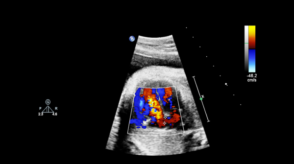
Fetal echocardiography is a specialized ultrasound test that provides a detailed view of your unborn baby’s heart its structure, function, and rhythm. Typically performed between the 18th and 24th weeks of pregnancy, this test allows fetal medicine specialists in Amritsar to identify potential heart abnormalities, ranging from congenital heart defects to arrhythmias or irregular heart rhythms.
By detecting these conditions early, doctors can prepare for necessary interventions during or immediately after birth and in some cases, even treat the condition before delivery.
When is Fetal Echocardiography Recommended?
- There’s a family history of congenital heart disease
- You’ve previously had a child with heart defects
- Genetic concerns are identified during screening
- You’ve taken certain medications (e.g., for seizures or depression) during pregnancy
- There has been drug or alcohol use during pregnancy
- The mother has conditions like diabetes, lupus, or autoimmune disorders
- Any irregularities are noticed during a routine ultrasound
- The pregnancy is a result of assisted reproductive technologies (IVF, IUI, etc.)
Types of Fetal Echocardiography:
-
Abdominal Echocardiography:
Similar to a regular ultrasound, the probe is placed on the mother’s abdomen to assess the baby’s heart. -
Transvaginal Echocardiography:
A small probe is gently inserted into the vagina for a closer view, especially useful in early pregnancy.
Both procedures are safe, non-invasive, and do not have any risk to the mother or baby. If the results are unclear or further evaluation is needed, your doctor may recommend additional imaging or diagnostic tests to confirm a diagnosis.
Visit leading fetal echocardiography test centers in Amritsar to get expert screening and timely insights into your baby’s heart health.
Frequently Asked Question
It can identify congenital heart defects, valve issues, structural abnormalities, and heart rhythm problems like arrhythmias.